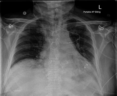
lv epicardial lead | epicardial lv lead implantation lv epicardial lead Between March 2008 and May 2014 an epicardial LV lead was implanted in 32 . Crocodile skin, Leather, Ostrich skin. Bracelet color. Black, Blue, Bordeaux, Brown, Red. Clasp material. Gold/Steel, Silver, Steel, Yellow gold. Find low prices for 26 Cartier ref. Cartier Must de Tank watches on Chrono24. Compare deals and buy a ref. Cartier Must de Tank watch.
0 · transthoracic introduction epicardial lead
1 · transthoracic epicardial lead placement
2 · transthoracic epicardial lead insert
3 · left ventricular lead frontiers
4 · invasive epicardial lead placement
5 · epicardial lv lead replacement
6 · epicardial lv lead placement
7 · epicardial lv lead implantation
$18K+
Left ventricular (LV) lead positioning remains an important variable that predicts the response to CRT. Anatomical and technical challenges can hinder optimal LV lead placement using traditional lead implantation approaches.An optimal placement of the left ventricular (LV) lead appears crucial for the .Between March 2008 and May 2014 an epicardial LV lead was implanted in 32 .Epicardial LV lead positioning has the advantage of direct visualization and .
Therefore, epicardial LV lead implantation using VATS can be effectively and .An optimal placement of the left ventricular (LV) lead appears crucial for the intended hemodynamic and hence clinical improvement. A well-localized target area and tools that help .
LV Lead Location and Baseline Clinical Characteristics. The LV lead position was assessed in 799 patients (55% patients ≥65 years of age, 26% female, 10% LVEF ≤25%, 55% ischemic cardiomyopathy, and 71% LBBB) .Minimally invasive left ventricular epicardial lead placement is safe and effective, offering selection of the best pacing site with minimal morbidity; it can be considered a primary option for resynchronization therapy. Between March 2008 and May 2014 an epicardial LV lead was implanted in 32 patients after failed transvenous LV lead placement using a left-sided lateral minithoracotomy .
The present article reviews the literature on image-guided cardiac resynchronization therapy (CRT) studies. Improved outcome to CRT has been associated with the placement of a left ventricular (LV) lead in the latest . Epicardial LV lead positioning has the advantage of direct visualization and selection of the most suitable surface of LV, also avoiding areas of epicardial fat or fibrosis that .
Using an epicardial lead placed on the LV free wall via thoracotomy and endocardial leads placed in the right atrium (RA), left atrium (LA) via the coronary sinus (CS) . Therefore, epicardial LV lead implantation using VATS can be effectively and safely used as a rescue method for patients with recurrent LV lead dislodgement, or even as a .Left ventricular (LV) lead positioning remains an important variable that predicts the response to CRT. Anatomical and technical challenges can hinder optimal LV lead placement using traditional lead implantation approaches.
An optimal placement of the left ventricular (LV) lead appears crucial for the intended hemodynamic and hence clinical improvement. A well-localized target area and tools that help to achieve successful lead implantation seem to be of utmost importance to . LV Lead Location and Baseline Clinical Characteristics. The LV lead position was assessed in 799 patients (55% patients ≥65 years of age, 26% female, 10% LVEF ≤25%, 55% ischemic cardiomyopathy, and 71% LBBB) with a follow-up of 29±11 months.Minimally invasive left ventricular epicardial lead placement is safe and effective, offering selection of the best pacing site with minimal morbidity; it can be considered a primary option for resynchronization therapy.
We retrospectively assessed two types of sutureless screw-in left ventricular (LV) leads (steroid eluting vs. non-steroid eluting) in cardiac resynchronization therapy (CRT) implantation with regards to their electrical performance. Between March 2008 and May 2014 an epicardial LV lead was implanted in 32 patients after failed transvenous LV lead placement using a left-sided lateral minithoracotomy or video-assisted thoracoscopy (mean age 64 ± 9 years). The present article reviews the literature on image-guided cardiac resynchronization therapy (CRT) studies. Improved outcome to CRT has been associated with the placement of a left ventricular (LV) lead in the latest activated segment free from scar. Epicardial LV lead positioning has the advantage of direct visualization and selection of the most suitable surface of LV, also avoiding areas of epicardial fat or fibrosis that can cause increase in pacing thresholds.
Using an epicardial lead placed on the LV free wall via thoracotomy and endocardial leads placed in the right atrium (RA), left atrium (LA) via the coronary sinus (CS) and RV, they demonstrated a decrease in pulmonary capillary wedge pressure and an increase in cardiac output with temporary four-chamber pacing.
Therefore, epicardial LV lead implantation using VATS can be effectively and safely used as a rescue method for patients with recurrent LV lead dislodgement, or even as a de novo way for those with unfavorable cardiac vein anatomy or when better targeted LV pacing is .Left ventricular (LV) lead positioning remains an important variable that predicts the response to CRT. Anatomical and technical challenges can hinder optimal LV lead placement using traditional lead implantation approaches.
An optimal placement of the left ventricular (LV) lead appears crucial for the intended hemodynamic and hence clinical improvement. A well-localized target area and tools that help to achieve successful lead implantation seem to be of utmost importance to . LV Lead Location and Baseline Clinical Characteristics. The LV lead position was assessed in 799 patients (55% patients ≥65 years of age, 26% female, 10% LVEF ≤25%, 55% ischemic cardiomyopathy, and 71% LBBB) with a follow-up of 29±11 months.Minimally invasive left ventricular epicardial lead placement is safe and effective, offering selection of the best pacing site with minimal morbidity; it can be considered a primary option for resynchronization therapy.
transthoracic introduction epicardial lead
We retrospectively assessed two types of sutureless screw-in left ventricular (LV) leads (steroid eluting vs. non-steroid eluting) in cardiac resynchronization therapy (CRT) implantation with regards to their electrical performance. Between March 2008 and May 2014 an epicardial LV lead was implanted in 32 patients after failed transvenous LV lead placement using a left-sided lateral minithoracotomy or video-assisted thoracoscopy (mean age 64 ± 9 years). The present article reviews the literature on image-guided cardiac resynchronization therapy (CRT) studies. Improved outcome to CRT has been associated with the placement of a left ventricular (LV) lead in the latest activated segment free from scar.
Epicardial LV lead positioning has the advantage of direct visualization and selection of the most suitable surface of LV, also avoiding areas of epicardial fat or fibrosis that can cause increase in pacing thresholds.
Using an epicardial lead placed on the LV free wall via thoracotomy and endocardial leads placed in the right atrium (RA), left atrium (LA) via the coronary sinus (CS) and RV, they demonstrated a decrease in pulmonary capillary wedge pressure and an increase in cardiac output with temporary four-chamber pacing.

real prada vs fake prada
aaa replica prada sneakers
Cartier’s solution was the introduction of the Cartier Tank Américaine. Bigger, bolder yet still possessing the desirable curved case, two versions of the Tank Américaine launched in the late 80s with little fanfare.
lv epicardial lead|epicardial lv lead implantation


